Medial Geniculate Body
This oval body is placed underneath pulvinar of thalamus lateral to superior colliculus. Inferior colliculus is connected to medial geniculate body by a band known as inferior brachium. Medial geniculate body is the diencephalic relay station of cochlear pathway for hearing. So afferent fibers reach this nuclear mass coming from below and efferent fibers go upto the auditory cortex.
Afferent: These are narrow compact ascending fiber bundle called lateral lemniscus which are axons of nerve cells from superior olivary nucleus and nucleus of trapezoid body in lower end of pons. Some of the fibers pass to medial geniculate body after relaying in inferior colliculus. Beyond inferior colliculus, fibers enter medial geniculate body through inferior brachium.
Efferent: Efferent fibers are axons of nerve cells in medial geniculate body. These fibers form auditory radiation. These form sublentiform part of internal capsule to end in auditory cortex which is present in the form of transverse gyri on upper surface of superior temporal gyrus (Area 41 and 42).
Lateral Geniculate Body
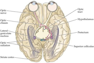
This is positioned also underneath pulvinar of thalamus and lateral to medial geniculate body and smaller in size. It is connected to superior colliculus of midbrain by superior brachium. Lateral geniculate body is the diencephalic relay station of visual pathway.
Lateral geniculate body of one side receives visual information of ipsilateral half (right or left) of both retina, so form contralateral half of field of vision of both eyes.
Afferent: Neurons of lateral geniculate body are arranged in six layers which are numbered one to six from ventral to dorsal aspects. Afferent fibers reach lateral geniculate body via optic tract. The axons of multipolar ganglionic neurons of retina leave eyeball through optic nerve, optic chiasma and then through optic tract to relay in lateral geniculate body. Lateral geniculate body receives almost all the fibers of optic tract except some, which go to pretectal nucleus for light reflex. It is known that lateral geniculate body of one side receive fibers from same half (right or left) of both retina. Laminae 1, 4 and 6 of lateral geniculate body receive fibers of retina of opposite side and laminae 2, 3 and 5 receive fibers of retina of same side.
Efferent: Efferent fibers from all the layers of lateral geniculate body form geniculocalcarine tract. It is also known as optic radiation which is the thalamocortical (corticopetal) component of posterior thalamic radiation. These fibers pass through retrolentiform part of internal capsule.
Source: Easy and Interesting Approach to Human Neuroanatomy (Clinically Oriented) (2014)