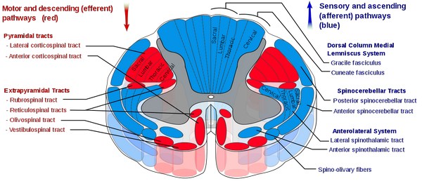
General consideration: These tracts are two in number, called ventral (anterior) and dorsal (posterior) spinocerebellar tracts. Instead of going upto sensory area of cerebral cortex via thalamus, they terminate in cerebellar cortex.
Functions of spinocerebellar tracts are concerned wih coordination of movements.Impulse is carried from neuromuscular spindle (muscle spindle), neurotendinous spindle (Golgi tendon organ) and joint receptors.
End organs are stimulated due to stretching of muscles and tendons, and movements of the joints. Both the spinocerebellar tracts are situated in the form of narrow strip covering the peripheral part of lateral funiculus, being anteroposteriorly related. Both the tracts are ipsilateral. But it is important to note that fibers for ventral spinocerebellar tractcross at the level of corresponding spinal cord segments. But for the second time the tract crosses as a whole at the level of midbrain.
Both the tracts are made of myelinated fibers of large diameter. Ventral spinocerebellar tract also contains some thin calibered fibers. Individual characteristics of either of the tracts will be clear from their description below.
Dorsal spinocerebellar tract
It is formed by axons of Clerke’s column of cells. Therefore this tract start formation and so also starts ascending from second or third lumbar segment of spinal cord. It is also not difficult to understand that dorsal spinocerebellar tract gets formed from L2 / L3segment to T1 segment. It receives therefore input from the trunk through T1–L2 / L3 segmental spinal nerve. It is interesting to note that it also receives inputs from lower limb. Proprioceptive impulse from neuromuscular spindle, neurotendinous spindle and joints of lower limbs are carried by dorsal column (fasciculus gracilis). Reaching upto L2 / L3 segments, collaterals are given from dorsal column to relay in Clarke’s column of cells of L2/L3 segments.
These collateral are given by the fibers of dorsal column carrying impulse from the lower limb through spinal segment, as they reach the level of L2/L3 segments. Again above T1 segment, proprioceptive sensations from neuromuscular spindle, neurotendinous spindle and joint receptors ascends through fasciculus cuneatus to relay also in accessory cuneate nucleus which is a smaller oval bulge superolateral to nucleus cuneates. Fibers from this nucleus reach the cerebellum via cuneocerebellar tract.
Ventral spinocerebellar tract
Ventral (anterior) spinocerebellar tract is formed by axons of tract neurons of lamina V to VII of spinal cord in addition to Clarke’s column of cells.These second order neurons, receives nformation from muscle, tendon and joint receptors via the first order neurons, which are primary sensory neurons of posterior root ganglia. In addition, ventral spinocerebellar tract carries sensory information from skin and subcutaneous tissue also. These sensation are carried via ventral spinocerebellar tract from trunk as well as upper and lower limb.
Before the tract is being formed by the axons of lamina V to VII and also Clarke’s column of cells, majority of fibers cross the midline along ventral white commissure of spinal cord, while the minority of fibers remain in same side. Fibers take the position over a narrow strip of anterior peripheral part of lateral funiculus to ascend upwards in front of dorsal spinocerebellar tract. Fibers of ventral spinocerebellar tract run upwards carrying contralateral fibers, through the brainstem beyond spinal cord. Reaching the level of midbrain, fibers cross the midline for second time to reach cerebellar hemisphere of the same side through superior cerebellar peduncle.
Source: Easy and Interesting Approach to Human Neuroanatomy (Clinically Oriented) (2014)