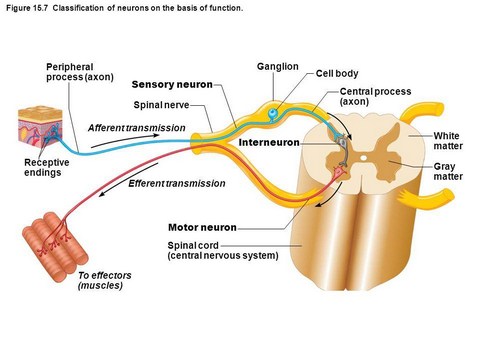
- Tract neurons: These are sensory or afferent neurons present in posterior horn. They are so called because their axons form compact bundles of ascending (sensory) tracts to relay in higher centers in brain.
They receive synaptic connections from central process of pseudounipolar neurons of posterior root ganglion which collect sensory informations from peripheral sensory end organs (receptors). It is important to note at this stage that axons of tract cells may ascend in the same side or may cross the midline and then ascend along opposite side of spinal cord to form uncrossed (ipsilateral) or crossed (contralateral) tracts respectively. - Motor neurons (efferent neurons): The neurons of anterior horn are motor neurons. Their axons, leaving spinal cord through ventral root, end in voluntary muscles via spinal nerve.terminate in extrafusal fibers of voluntary muscles, stimulation of which results in muscular contraction.
These motor neurons of spinal cord are called lower motor neurons on which relay the axons of nerve cells situated at higher centers (brain) which are called upper motor neurons. Motor neurons of anterior horn of spinal cord sending axons to voluntary muscles are of two types:
- Alpha motor neurons: Their cell bodies are more than 25 microns in size and their axon
- Gamma motor neurons: Cell bodies of these neurons are less than 25 microns in size and their axons terminate in intrafusal fibers of voluntary muscles, stimulation of which is concerned with increase in muscle tone.
- Interneurons (internuncial neurons): These are example of short axoned Golgi type II neurons.
Their axon as well as dendrite are shorter being confined in the gray matter of spinal cord. Functionally they are interconnecting in nature forming synaptic link between sensory and motor neuron which together form a local reflex arc. Internuncial neuron also leads to an advantage by connecting one first order of neuron, through its multiple axon collateral, to the multiple third order of neurons.
Further classification of motor and sensory neurons:
It is already understood from the knowledge of embryological background that, mantle zone of developing spinal cord forming gray matter forms four column of cells which are as follows from ventral to dorsal aspect.
- Somatic motor (efferent)
- Visceral motor (efferent)
- Visceral sensory (afferent)
- Somatic sensory (afferent).
- T1 – L2 segments of spinal cord: Here both the motor and sensory cell groups form additional horns called intermediolateral horn, where visceral efferent and visceral afferent cell groups form motor and sensory centers of sympathetic part of autonomic nervous system respectively.
- S2, S3 and S4 segments of spinal cord: Here the cell groups are present in intermediate area of gray matter without forming any additional lateral horn. Visceral efferent and visceral afferent neurons in these cell groups form motor and sensory centers of parasympathetic part of autonomic nervous system respectively.
Source: Easy and Interesting Approach to Human Neuroanatomy (Clinically Oriented) (2014)