Internal structure of spinal cord can be understood through the study of its cross section.
Cross section of spinal cord shows fundamentally following two components.
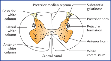
- Central gray matter: This is so called due to grayish color of cell bodies of neurons. Central zones of gray matter looks like ‘butterfly’ on cross section. Roughly it resembles the capital letter ‘H’. Intermediate bar of ‘H’ represents the body, whereas wings of butterfly are represented by two limbs of the letter. Basic components of spinal gray matter: Intermediate part of spinal gray matter is traversed centrally be central canal of spinal cord throughoutits whole length.
Central canal of spinal cord is lined by ependymal cells. The gray matter anterior and posterior to central canal are known as anterior and posterior gray commissures respectively. Each side of spinal cord gray matter is composed of following components:- Anterior known as anterior horn
- Intermediate area
- Posterior known as posterior horn.
When considered the whole length of spinal cord, anterior gray horn forms the anterior gray column and posterior gray horn forms the posterior gray column. In addition to the above mentioned three components, first thoracic to second lumbar segments (T1 – L2) of spinal cord gray matter show a lateralprojection of intermediate area, which is known as intermediolateral cell column. Neurons of this area constitute sympathetic center of autonomic nervous system. It is important to note at this stage that spinal center of parasympathetic nervous system is formed by neurons of intermediate area of second, third and fourth (S2, S3 and S4) sacral segments of spinal cord. The different components so also the entire gray matter of spinal cord show variations in appearance in different regions of spinal cord, because it depends upon the relative amount of nerve cells. Basically gray matter is proportionately broader in lower cervical and lumbosacral regions of spinal cord. - Peripheral white matter: This is mainly made up of compact bundles of nerve fibers running vertically either in ascending or in descending direction.
These fibers in the bundles are myelinated. The lipid-protein substance of myelin sheath of nerve fibers is white in color for which this peripheral zone of spinal cord is called white matter.
The bundles of ascending fibers carry sensory informations to the centers of brain above the level of spinal cord. The descending bundles carry impulse from higher motor centers of brain (above spinal cord) to the motor neurons situated in anterior horn of spinal cord.
On either side of midline, the white matter is composed of following three components called Funiculi (Singular – Funiculus).
- Anterior funiculus: It is the part of white matter between anterior median fissure and anterolateral sulcus presenting outgoing fibers of ventral nerve root. Anterior funiculi of two sides are bridged by a thin midline strip of white matter known as anterior white commissure.
- Lateral funiculus: It is the part of white matter demarcated between outgoing fibers of ventral root and incoming fibers of dorsal root of spinal nerve.
- Posterior funiculus: It is the part of white matter between posterior median sulcus and incoming fibers of dorsal root of spinal nerve attached to the posterolateral sulcus. Posterior funiculi of both sides are separated incompletely or even completely by posterior median septum.
The bundles of fibers either ascending (sensory or afferent) or descending (motor or efferent) are called tracts or fasciculi (Singular-Fasciculus). It is interesting to note at this stage that posterior funiculus is composed of only ascending tracts whereas anterior and lateral funiculi are composed of both ascending as well as descending tracts.
All neurons of spinal cord are multipolar. Fundamentally the three different zones of spinal gray matter are made up of following four different neuronal groups.
- Posterior horn: Sensory (afferent ) or tract neurons
- Anterior horn: Motor (efferent) neurons.
- Intermediate area:
- Interconnecting neurons (interneurons), and
- Parasympathetic neurons at S2, S3 and S4 segments only.
- Intermediolateral area: Sympathetic neurons (only T1 – L2 segmetns). Both the sympathetic as well as parasympathetic areas are composed of motor and sensory neurons.
Source: Easy and Interesting Approach to Human Neuroanatomy (Clinically Oriented) (2014)