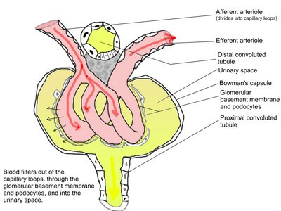
The first step in the formation of urine is filtration at the glomerulus. Glomerulus is a tufted network of capillaries, enclosed in the Bowman’s capsule. The renal plasma filters through the capillary endothelium and enters the Bowman’s capsule. The ultra structure of the glomerulus, reveals, the presence of three layers for the filtration barrier. They are:
- Capillary endothelial layer
- Basement membrane
- Visceral layer of Bowman’s capsule.
The capillary endothelial cells have fenestrae with 8 nm size. The solutes pass through this, depending on their molecular size and their electrical charge. The basement membrane is made of proteoglycan material, which acts as a sieve, allowing the solutes to easily pass through.
The visceral layer of the Bowman’s capsule is called the podocytes, as the apical border shows the presence of foot like processes, which encircle the endothelium. There are slits or gaps between the foot like processes, through which, water and solutes filter through and enter the lumen. The glomerular membrane per se includes all these three layers for filtration.
There are mesangial cells present between the capillary loops and between basement membrane and capillaries. These cells show contractile function. It can cause obliteration of the capill ary lumen, reducing the renal blood flow and GFR. Agents like angiotensin II, vasopressin, etc, can cause mesangial cell contraction. Mesangial cells are also present outside the glomerulus and they are called extraglomerular mesangial cells.
They are present in the JGA. The filtration of solutes depends on their size and electrical charge. Anions up to 4 nm show filtration and at 4 nm size, the filtration is only 50% of the neutral substances of same size. The anions are negatively charged and the cell membrane also has sialoproteins, which are negatively charged. The two negatively charged particles repel each other and that’s why, albumin, which is around 7 nm is not filtered, eventhough, the pore size of the endothelium is slightly greater than 7 nm. When glomerular membrane is damaged by inflammation or infections, i.e., glomerulonephritis, the negative charge on the membrane is lost and allows albumin to appear in the urine.
Source: Textbook of Physiology, 3E (Chandramouli) (2010)