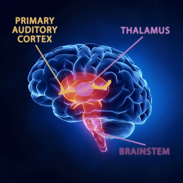
- Thalamus is the largest component of diencephalon.
- It is an egg-shaped ovoid mass of gray matter, one on either side of midline.
- A narrow midline cleft between thalami of two sides is third ventricle of brain.
- Gray mass of thalamus forming different nuclei forms the cell stations for all contralateral sensory pathways of body except the olfactory pathway.
- Two thalami are interconnected by a compact band of white matter crossing midline. It is called interthalamic adhesion.
- It is the thalamus component of dorsal diencephalon, which merges with the two components of ventral diencephalon as follows.
- In anterior plane: Merges with hypothalamus.
- In posterior plane: Merges with subthalamus.
- Ovoid mass of thalamus is anteroposteriorly elongated with following dimensions.
Anteroposterior measurement – 3.5 cm
Transverse measurement – 1.5 cm - Longer anteroposterior axis is directed forwards and medially.
Poles
Anterior pole is narrower and more close to the midline. It forms posterior boundary of interventricular foramen of Monro. Posterior pole is broader. It is known as Pulvinar. Pulvinars of both side are separated by a narrow interval which lodges pineal gland. Pulvinar is the part of thalamus which overhangs lateral geniculate body of metathalamus.
Surfaces
- Superior surface: It forms the floor of central part or body of lateral ventricle along with thalamostriate vein, stria terminalis and body of caudate nucleus which are mediolaterally placed. Superior surface of thalamus is covered by thin layer of white matter called stratum zonale.
- Medial surface: It forms lateral boundary of third ventricle. Medial surfaces of both thalami are connected by interthalamic adhesion.
- Lateral surface: It is immediately covered by a thin lamina of white matter called external medullary lamina. Beyond this, lateral surface is related to thick compact fibrous band of posterior limb of internal capsule. Superior and medial surfaces are covered by ependyma lining the cavity of lateral ventricle and third ventricle respectively.
Source: Easy and Interesting Approach to Human Neuroanatomy (Clinically Oriented) (2014)