- Olfactory receptors (neuroreceptors) present in specialized area of nasal mucosa and olfactory nerves.
- Two orders of neurons.
- Olfactory area of cerebral cortex present in temporal lobe.
End organs or receptors for olfactory pathway are specialized neurons present in specialized area of mucous membrane of nasal cavity called olfactory epithelium.
Olfactory epithelium
This epithelium lines uppermost part mucous membrane of nasal cavity which is:
- Uppermost part of lateral wall of nose along with sphenoethmoidal recess above the level of superior nasal concha.
- Uppermost part of nasal septum (medial wall of nose), which is formed by perpendicular plate of ethmoid bone.
- Roof of the nose between above mentioned lateral and medial walls.
- Receptor cells: Which are specialized bipolar neurons.
- Supporting cells: These are tall columnar interstitial cells which intervenes between receptor cells having supportive function.
- Basal cells: These are shorter cells resting on basement membrane intermingled with other cells. Basal cells are progenitor cells concerned with replacement (renewal) of receptor cells.
Olfactory receptor cells are specialized bipolar neurons scattered among supporting cells. Peripheral processes of these bipolar cells are wider and extend to the surface of nasal mucous membrane. Form the end of peripheral process, a number of short cilia arise which project into the mucus covering olfactory mucous membrane. These are called olfactory hairs. These projecting olfactory hairs react to odors of inhaled air and stimulates olfactory receptor cells.
Central processes of olfactory receptors are finer which form olfactory nerve fibers. These finer fibers aggregate to form 20 bunches. These are nonmyelinated. These 20 bunches of finer nonmyelinated fibers form olfactory nerves. It is clear therefore, unlike other cranial nerve, in each side, olfactory nerve is multiple in number.
First order of neurons
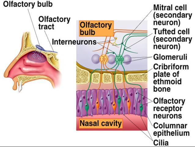
Bunch of olfactory nerve, the central processes of olfactory receptor cells, pass upwards from roof nose through foramina in cribriform plate of ethmoid bone to reach anterior cranial fossa. Reaching anterior cranial fossa, olfactory nerves terminate in first order of neurons of olfactory pathway which are present inside an ovoid flattened structure lodged on orbital surface of frontal lobe of cerebrum. It is called olfactory bulb. Neurons present inside olfactory bulb are of following types.
- Mitral cells
- Tufted cells
- Granule (Stellate) cells.
Mitral cells are largest cells in olfactory bulb. Incoming fibers of olfactory nerve form synaptic connections with mitral cells. These synaptic junctions also receive connection from tufted cells and granule cells. Junctional areas of these cells with olfactory nerve ending are known as glomeruli. Axons of these cells, within olfactory bulb, which are 1st order of neurons, pass backward to be continued as olfactory tract.
n Olfactory tract: It is a narrow and flat band of white matter extending from olfactory bulb to run backwards along olfactory sulcus on orbital surface of frontal lobe of cerebrum. Olfactory tract passes backward upto anterior perforated substance of base of the brain where it divides in an angular fashion into lateral and medial olfactory striae. Anterior perforated substance is embrassed anterolaterally and anteromedially by lateral and medial olfactory striae. The two straie form olfactory trigone.
Medial olfactory stria carries axons from cells of olfactory bulb which cross the midline as a component of anterior commissure to pass to olfactory bulb of opposite side. Lateral olfactory stria carries axons of 1st order of neurons present in olfactory bulb to the primary olfactory area beyond anterior perforated substance.
Intermediate olfactory stria is a short band of fibers, being occasionally present, passes from the angle of olfactory trigone to a small elevation on anterior perforated substance which is called olfactory tubercle.
Source: Easy and Interesting Approach to Human Neuroanatomy (Clinically Oriented) (2014)