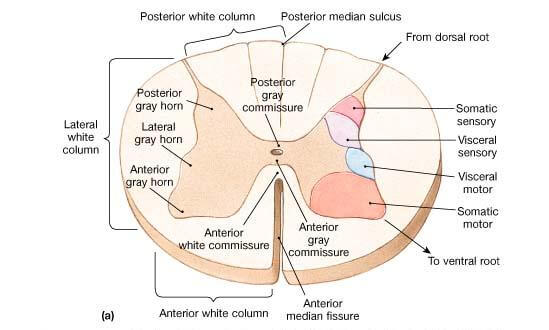
From apex towards the base they are as follows:
- Nucleus marginalis: It is ill-defined, thin strip of gray matter extending throughout whole length of spinal cord. It contains neurons of varying size with intermingling reticulum of fibers.
- Substantia gelatinosa of Rolando: It is a narrow area extending throughout the whole length of spinal cord. Afferent fibers, entering through posterior nerve root which carry pain and temperature sensations from synaptic connection with these neurons. Axons of these neurons cross midline and ascend through contralateral lateral white column of spinal cord to form lateral spinothalamic tract.
- Nucleus proprius: It is the main bulk of neurons in posterior horn and extend throughout wholelength of spinal cord. Nucleus proprius consists of following neurons.
- Some of these cells receive incoming posterior nerve root fibers which carry sensations of crude touch and pressure. However it is told these days that crude touch and pressure fibers end in many cell group of posterior horn.
- Axons of some of cells of nucleus proprius link adjacent segments of spinal cord for intraspinal coordination.
- Nucleus dorsalis (Clarke’s column): It is also called nucleus thoracis. It extends from eighth cervical to third or fourth lumbar segments of spinal cord. Nucleus dorsalis or Clarke’s column of cells are situated on medial side of base of dorsal gray horn and show a projection on posterior white column of spinal cord. Neurons of this column of varying size and shape are of following two varieties.
- Neurons which receive incoming afferent fibers via nerve root carrying unconscious proprioceptive sensation from muscle spindle and Golgi tendon organs. These neurons send axons which ascend through marginal strip of lateral white column forming dorsal as well as anterior spinocerebellar tract.
- Interneurons of Golgi type II characterized by short dendrites as well as short axons.
- Visceral afferent cell column: These cell groups are situated lateral to Clarke’s column of cells at the base of dorsal gray horn. But it exists in following two levels of spinal cord.
- T1 to L1/L2 segments of spinal cord: The cell groups receive sympathetic afferent fibers which enter the spinal cord through posterior root of spinal nerve. It receives sensations from wall of viscera.
- S2, S3 and S4 segments of spinal cord: These cell groups receive parasympathetic afferent fibers which also enter the spinal cord through posterior root of spinal nerve. It receives parasympathetic sensation from the wall of the viscera wherefrom carried via pelvic splanchnic nerve.
Source: Easy and Interesting Approach to Human Neuroanatomy (Clinically Oriented) (2014)