Structural characteristics
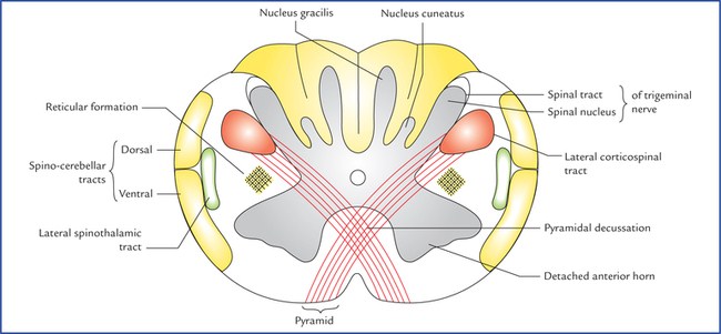
- At this level structure of medulla oblongata is almost similar to the structure of spinal cord, with centrally positioned gray matter and peripheral white matter.
- Ventral horn of gray matter gets separated from main mass due to decussation of pyramidal tract fibers which pass backwards and laterally to approach lateral white column before passing downwards to the spinal cord.
Gray matter:
- Central gray matter is traversed by more dorsally pushed central canal lined by ependyma.
- Apex of posterior horn of spinal cord is represented at this level by nucleus of spinal tract of trigeminal nerve. On either side it is directed backwards and laterally with further abduction.
- Medial to nucleus of spinal tract of trigeminal nerve, gray matter shows, on either side, two small bulge of gray matter, nucleus gracilis (medial) and nucleus cuneatus (lateral) which receive the fibers of fasciculus gracilis and fasciculus cuneatus respectively, which are the ascending tracts in posterior column of white matter.
- Anterior gray horn becomes detached from main mass of gray matter by decussating fibers of corticospinal (pyramidal) tract.
- Supraspinal nucleus of first cervical nerve: It is the upward continuation of anterior horn cells of first cervical segments of spinal cord. Axons of these neurons pass downward and are distributed along the ventral root of first cervical nerve.
- Ascending nucleus: It is the upward continuation of spinal nucleus of accessory nerve which is continuous below up to fifth cervical segment of spinal cord. Above it is continuous with nucleus ambiguous.
- Anterior column: On either side of ventral median fissure, area of anterior white column mainly presents the bundle of pyramidal tract fibers which shows decussation of fibers at this level. Through anterior column, also traverse tectospinal tract, vestibulospinal tract, anterior spinothalamic tract.
- Lateral Column:
- Peripherally: Dorsal and ventral spinocerebellar tracts.
- Centrally:
- Lateral corticospinal tract which is formed at this level after decussation of fibers of pyramid.
- At the center of lateral white column, a scattered group of nerve cells intermingled with nerve fibers form brainstem reticular formation.
- Lateral spinothalamic tract.
- Posterior column: It present upward continuation of fasciculus gracilis and fasciculus cuneatus of posterior white column of spinal cord. As already mentioned earlier, these two tracts will relay in next order of neurons in nucleus gracilis and nucleus cuneatus which are seen to appear at this level of medulla oblongata, ventral to the corresponding tracts.
Source: Easy and Interesting Approach to Human Neuroanatomy (Clinically Oriented) (2014)