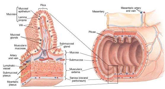
The digestive tract includes mouth, pharynx, esophagus, stomach, small intestine, large intestine, rectum and anus. The histological structure is mostly similar in all regions except esophagus and rectum. In these two regions, serous coat and attachment to peritoneum are not present.
The basic structure of digestive tract shows three layers namely from outwards, serous coat, middle muscular layer and inner mucous layer. The muscular coat is made of three layers namely, two layers of longitudinal and one layer of circular smooth muscle.
The wall of the GI tract has intrinsic nervous system, which consists of Auerbach’s plexus or myenteric plexus and Meissner’s plexus or submucous plexus. Between longitudinal and middle circular layer of smooth muscle, Auerbach’s plexus is present. These neurons are motor in nature and supply the smooth muscle. They also make connections with the neurons of Meissner’s plexus. The myenteric plexus is mainly concerned with controlling the motor activity of the gut.
Situated in the submucous layer (between middle circular layer of smooth muscle and inner mucous layer) there is another intrinsic nerve plexus called Meissner’s plexus. These neurons supply the secretory glands present in the wall of the gut. There is also interconnection between these two neuronal systems and together they are as enteric nervous system. The function of myenteric plexus is to produce peristalsis, by the contraction of the smooth muscle, whereas, the Meissner’s plexus is concerned with the sensing of osmolar changes, pH changes and chemical composition of food. In response to this stimulation, the Meissner’s plexus causes, secretion of digestive glands present in the wall of the gut. They are also sensitive to stretch, which is characteristic of these neurons.
The enteric nervous system action is independent of the extrinsic nerve supply to the gut. The extrinsic innervation can modify the activity of enteric nervous system. The extrinsic supply comes from autonomic nerves. The sympathetics give relaxation of smooth muscle and vasoconstriction of blood vessels, while, the parasympathetic (vagus and pelvic nerves) innervating the gut, causes contraction of smooth muscle, vasodilatation and secretion of digestive juices.
The actions of enteric nervous system are mediated through the secretion of transmitters. The released transmitters, besides, showing effects locally, act on surrounding cells (paracrine) and also on distant glands (endocrine) through circulation in the blood. In addition to norepinephrine, acetylcholine, serotonin, GABA secreting neurons, the enteric nervous system also has NO, substance P, VIP, somatostatin, CCK, GRP secreting neurons. NO secretion causes vasodilatation and relaxation of smooth muscle. VIP secreting neurons show, secretion of intestinal juice and relaxation of sphincters. The neurons secreting substance P, gives contraction of intestinal smooth muscle and stimulation of gastric acid secretion. The CCK secreting neurons show inhibition of the gastric motility and emptying, while somatostatin causes the inhibition of gastric and intestinal secretions.
Source: Textbook of Physiology, 3E (Chandramouli) (2010)