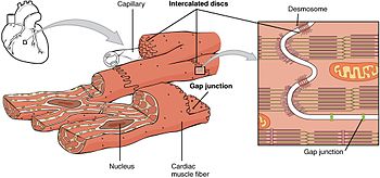
Cardiac muscle is a striated, involuntary muscle. The histology of the muscle shows, that it consists of interdigitating network of branches. The cell is cylindrical in shape, with all the cell organelles present in the cytoplasm. Each cell is separated from the adjoining cell by an intercalated disc. At the sides of these discs, there is a gap junction present. Through this, ions and electrical potentials can easily pass through, facilitating the spread of excitation to the entire myocardium.
Similar to skeletal muscle, there is presence of actin, myosin, troponin, and tropomyosin. There is also a well developed sarcoplasmic reticulum, and the triad is present at the Z line and not in the A–I junction, as in skeletal muscle. Cardiac muscle being an excitable tissue shows electrical properties, which gives rise to mechanical activity in the form of contraction. The mammalian heart has a pacemaker, from where the impulses originate. It is present at the sinoatrial node. This region is electrically more excitable than any other region of the heart. The resting membrane potential is low in this region, which is – 60 mv. There is decreased K+ exit in the previous repolarization of the action potential, which causes depolarization of the cell. The Ca++ channels open, especially the transient calcium channels to complete the prepotential. That is, the membrane potential falls from – 60 mv to – 40 mv. This phase is called prepotential.
At – 40 mv, the threshold of firing is reached, leading to opening of long lasting Ca++ channels. The influx of Ca++ through these channels produces the action potential.
Thus, we see that there is a spontaneous depolarization of the pacemaker cell due to the slow K+ exit. The completion of prepotential and formation of action potential are due to Ca++ influx and not by Na+ entry. Sympathetic stimulation increases the frequency of pacemaker potentials and hence increases heart rate. The vagal stimulation on the other hand, decreases the frequency or inhibits the formation of pacemaker potentials, depending on the intensity of vagal stimulation. This will give either decrease in the heart rate or complete inhibition of heart’s action.
Source: Textbook of Physiology, 3E (Chandramouli) (2010)