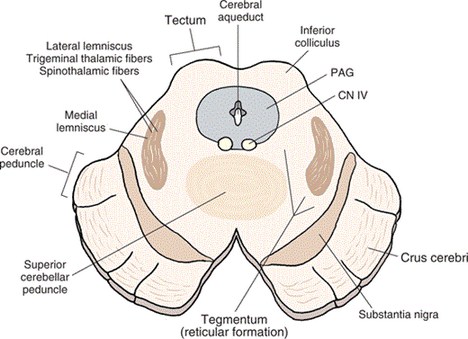
Central cavity of brainstem show different characteristics and names at different level. At lower end of medulla it is a narrow canal continuous below with central canal of spinal cord. At the level of pons and upper half of medulla oblongata, it becomes wide to form the cavity of 4th ventricle of brain. At the level of midbrain it is a narrow slit called aqueduct of Sylvius.
Fundamentally, neurons of basal plate are motor and those of alar plate are sensory in function. Throughout the whole length of developing brainstem, initially, many neurons of both basal as well as alar plate will form number of continuous columns of cells which are as follows:
In basal plate (from medial to lateral)
- Somatic efferent
- Branchial efferent (special visceral efferent)
- General visceral efferent.
- Somatic afferent
- Branchial afferent (special visceral afferent)
- General visceral afferent.
Migration of neurons of alar lamina: Apart from formation of sensory (afferent) nuclei of cranial nerves, neurons of alar plate will migrate from its original position either ventrally or further dorsally to form some other named nuclei in different level of brainstem (described below). This nuclei, as migrated, will intermingle with the components (white matter) of marginal zone.
Derivatives of marginal zone: It is already understood that, marginal zone is composed of processes of nerve cells of mantle zone. These processes will form different groups of bundles of nerve fibers which are basically of following two types:
- Vertical: These are either ascending (afferent) or descending (efferent) tracts of nerve fibers connecting spinal cord with various higher centers.
- Horizontal: These are fiber bundles connecting various centers of central nervous system with cerebellum in both direction, passing through 3 cerebellar peduncles.
- At the level of lower closed part of medulla oblongata: Cells of alar plate migrate further dorsally on either side of posterior median sulcus to form two nuclei.
- Medial: Nucleus gracilis
- Lateral: Nucleus cuneatus.
- At the level of upper half of medulla oblongata:
Cells of alar plate migrate ventrally in the peripheral plane of marginal zone in the form of
following nuclei.
Source: Easy and Interesting Approach to Human Neuroanatomy (Clinically Oriented) (2014)